Why are researchers looking at viruses?
T1D is partly influenced by genetic factors. However, genetic susceptibility alone cannot explain the rapidly increasing incidence of T1D worldwide. This has led researchers to investigate environmental factors that may trigger or accelerate the autoimmune process.
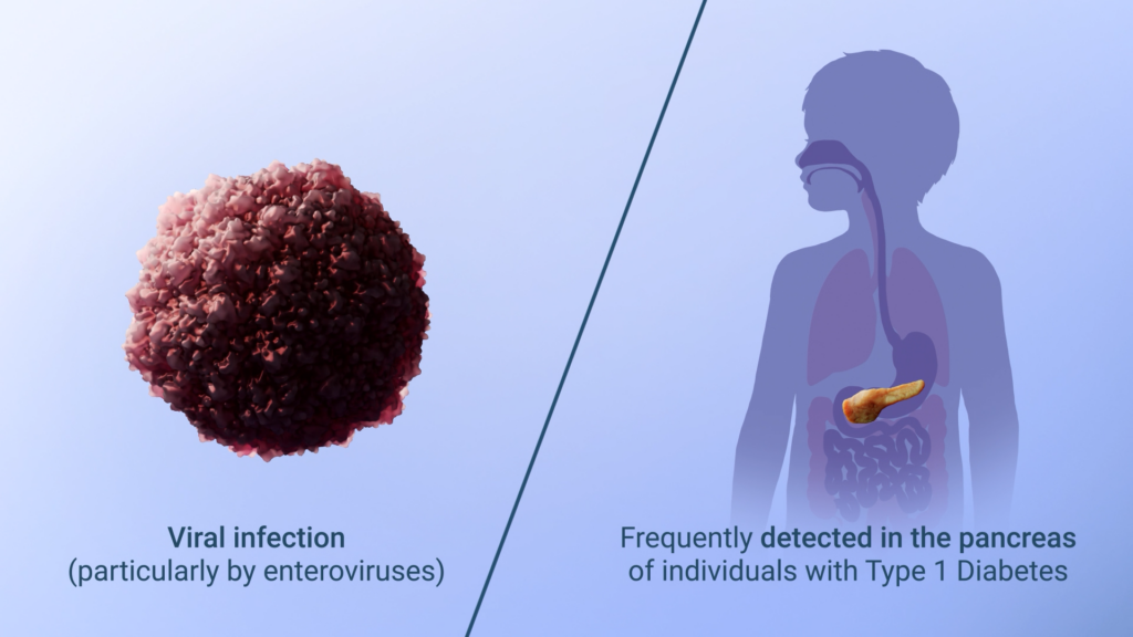
Among the most studied candidates are viral infections, particularly enteroviruses such as Coxsackie B viruses. A growing number of studies suggest that these viruses may play a key role in triggering T1D in genetically susceptible individuals.
Enteroviruses are common and usually cause mild illnesses. Even in the absence of severe symptoms the virus can enter the blood and spread to organs which are susceptible for the virus. Pancreas is once such organ, and beta-cells may be particularly susceptible since they express high amounts of molecules which the virus needs to enter the cell.
Interestingly, both enteroviral genetic material and live replicating viruses have been detected in pancreatic tissue from T1D patients, whereas viral presence has been clearly less common in healthy controls. Evidence of enterovirus infection has now been observed in both the early and later stages of the disease, strengthening the hypothesis that these viruses may contribute to the initiation and progression of T1D.

Diagram of pancreatic islet, showing insulin-producing beta cells and surrounding acini cells © Oslo University Hospital, 2026
How could a virus trigger type 1 diabetes?
By combining findings from studies investigating the link between enteroviruses and T1D, researchers have proposed a model describing how viral infection may lead to destruction of insulin-producing beta cells and eventually trigger autoimmune T1D.
The model, illustrated in the video presented here, begins with viruses entering beta cells in the pancreas and establishing a low-grade, persistent infection.
As the viruses slowly replicate over time, they induce cellular stress and promote the formation of misfolded proteins and neoantigens – altered molecules that the immune system may recognize as “non self”. This can initiate an autoimmune response, leading to the production of autoantibodies and ultimately, an immune-mediated attack of beta cells.